
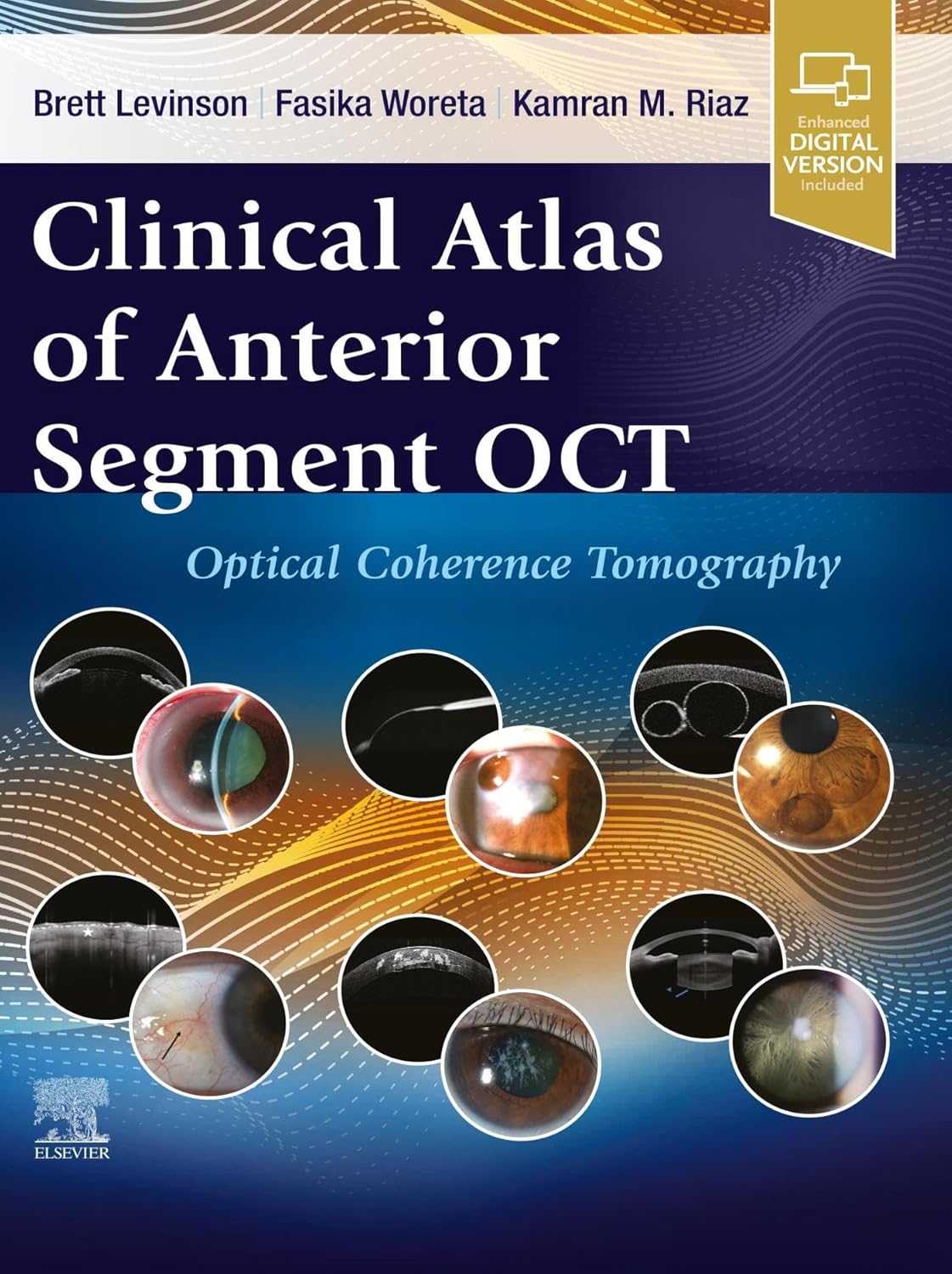
Discover the comprehensive world of optical coherence tomography (OCT) with our Clinical Atlas of Anterior Segment OCT. This expert guide is perfect for eye care providers needing to enhance their understanding of OCT uses, specifically in diagnosing and managing corneal and anterior segment conditions.
The Clinical Atlas of Anterior Segment OCT helps differentiate between various corneal pathologies, provides detailed anatomy of the angles, and serves as an invaluable tool for obtaining insights about the lens-capsule complex. It also aids in guiding contact lens fitting, among many other clinical applications.
Key features of our Clinical Atlas of Anterior Segment OCT include:
– An extensive coverage of the normal anatomy of the anterior segment and pathology of the conjunctiva, corneal epithelium, stroma and endothelium, lens, iris, anatomical angle, and more.
– Vital information on using AS-OCT in both clinical and surgical settings, including intraoperative AS-OCT.
– Over 500+ high-quality OCT images and clinical photos comparing normal anatomy and a wide range of pathologies, including common and rare disorders.
– Clear and easy-to-understand guidance on how to use, understand, and capture AS-OCT images.
– High-quality slit lamp images linked to the corresponding AS-OCT images, with clear labels to show the pathology side by side.
– Valuable clinical pearls in each chapter linking key AS-OCT and clinical findings to everyday practice.
Enhance your optical coherence tomography (OCT) skills and ensure accurate diagnosis and management of various eye diseases. With the Clinical Atlas of Anterior Segment OCT, you have a reliable and comprehensive guide to help you navigate through the intricate world of anterior segment diseases. Read more about this indispensable tool.
Authors:
Brett Levinson (Editor),
(Editor),
(Editor)
Edition:
Publication Date:
February 10, 2024
From the book:


Reviews
There are no reviews yet.