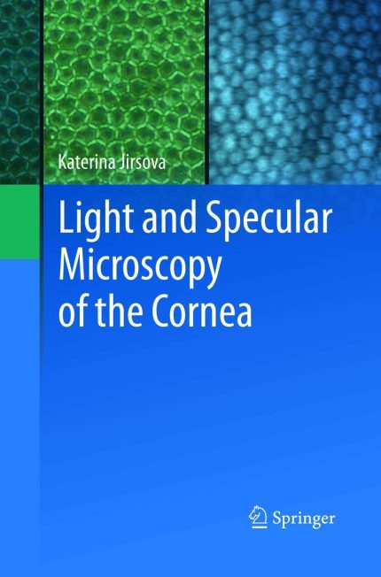
Introducing the comprehensive “Atlas of Light and Specular Microscopy of the Cornea,” a visual guide that offers valuable insights into the intricate world of corneal health and pathology. This atlas showcases a diverse range of photographs capturing the nuances of both healthy and diseased corneas, along with those primed for grafting procedures. Whether you are a medical professional, researcher, or simply intrigued by the fascinating realm of corneal anatomy, this resource is designed to enlighten and educate.
Key Features of the Atlas:
– Detailed imagery of the corneal layers: Explore the epithelium with its superficial and basal cells, the stroma housing keratocytes, and the crucial endothelium.
– In-depth analysis of corneas prepared for grafting: Focus on the endothelial layer and learn about the key factors influencing grafting eligibility, such as cell viability, polymeghatism, and stromal irregularities.
– Evaluation of stored corneal tissue: Witness the impact of tissue culture and hypothermic conditions on corneal suitability for grafting, providing a valuable resource for transplant assessments.
– Pathological insights: Delve into the world of corneal abnormalities through detailed photographs of pathological corneal explants, shedding light on various conditions and their visual manifestations.
By combining scientific rigor with visual clarity, this atlas serves as a valuable tool for understanding the complexities of corneal health and pathology. Whether you seek to enhance your knowledge base, aid in clinical decision-making, or simply appreciate the wonders of ocular anatomy, this resource is a must-have addition to your library.
Explore the “Atlas of Light and Specular Microscopy of the Cornea” today and embark on a visual journey through the intricate
Authors:
Katerina Jirsova (Author)
Edition:
Softcover reprint of the original 1st ed. 2017
Publication Date:
June 6, 2019
From the book:



Reviews
There are no reviews yet.