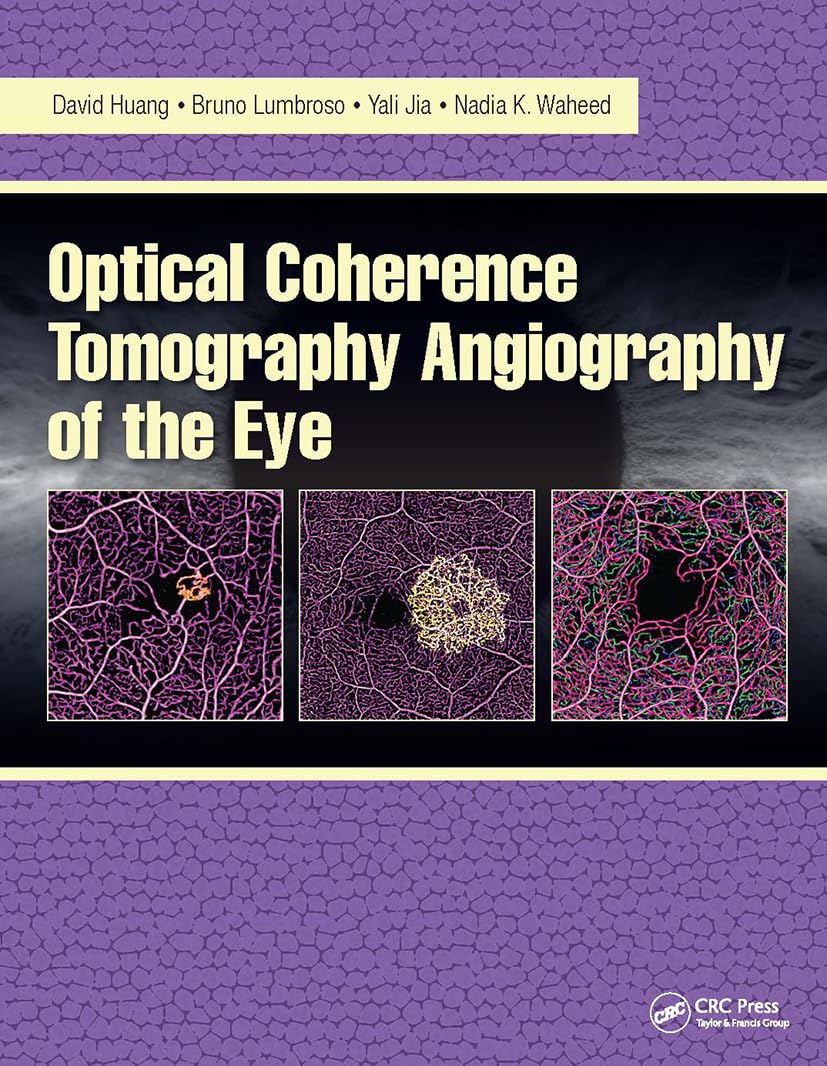
Optical Coherence Tomography (OCT) angiography is a breakthrough imaging technology taking the world of ophthalmology by storm. This innovative modality, which is being adopted by retina centers globally, offers key advantages over traditional dye-based fluorescein angiography (FA) and indocyanine green (ICG) angiography:
– OCT angiography uses motion as intrinsic contrast, eliminating the need for injecting any intravenous dye, making it a safer alternative.
– The technology employs infrared light that is invisible to the patient, ensuring a comfortable experience.
– It only requires a few seconds per scan, making it both user-friendly and patient-friendly.
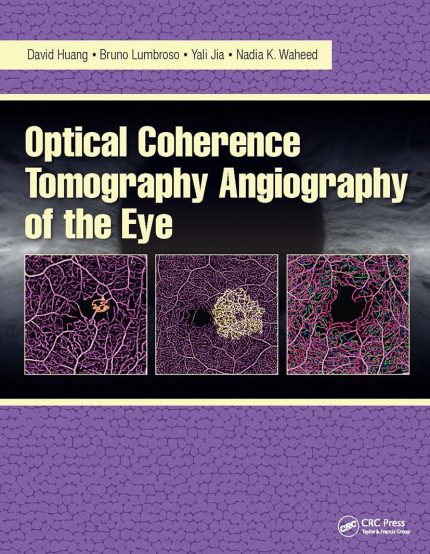
“Optical Coherence Tomography Angiography of the Eye,” a comprehensive guide by Drs. David Huang, Bruno Lumbroso, Yali Jia, and Nadia Waheed, provides in-depth information on the clinical application and essential principles of this advanced technology. Here’s what the book offers:
– A detailed overview of high-speed OCT systems and algorithms to extract flow contrast.
– Insightful explanation of OCT imaging of the normal eye and findings associated with various diseases.
– Practical tips on how to handle artifact and pitfalls.
– A deep-dive into the 3-dimensional aspect of OCT angiography, highlighting its ability to visualize difficult-to-image vascular beds like the 4 retinal vascular plexuses (radial peripapillary, superficial, intermediate, and deep), the choriocapillaris, and the deeper choroidal vessels.
Recognized for its noninvasive nature and simplicity, OCT angiography is rapidly carving a niche in daily ophthalmological practice, potentially replacing fluorescein angiography. “Optical Coherence Tomography Angi
Authors:
David Huang (Author),
(Author),
(Author),
(Author)
Edition:
1st
Publication Date:
July 17, 2017
From the book:

Reviews
There are no reviews yet.