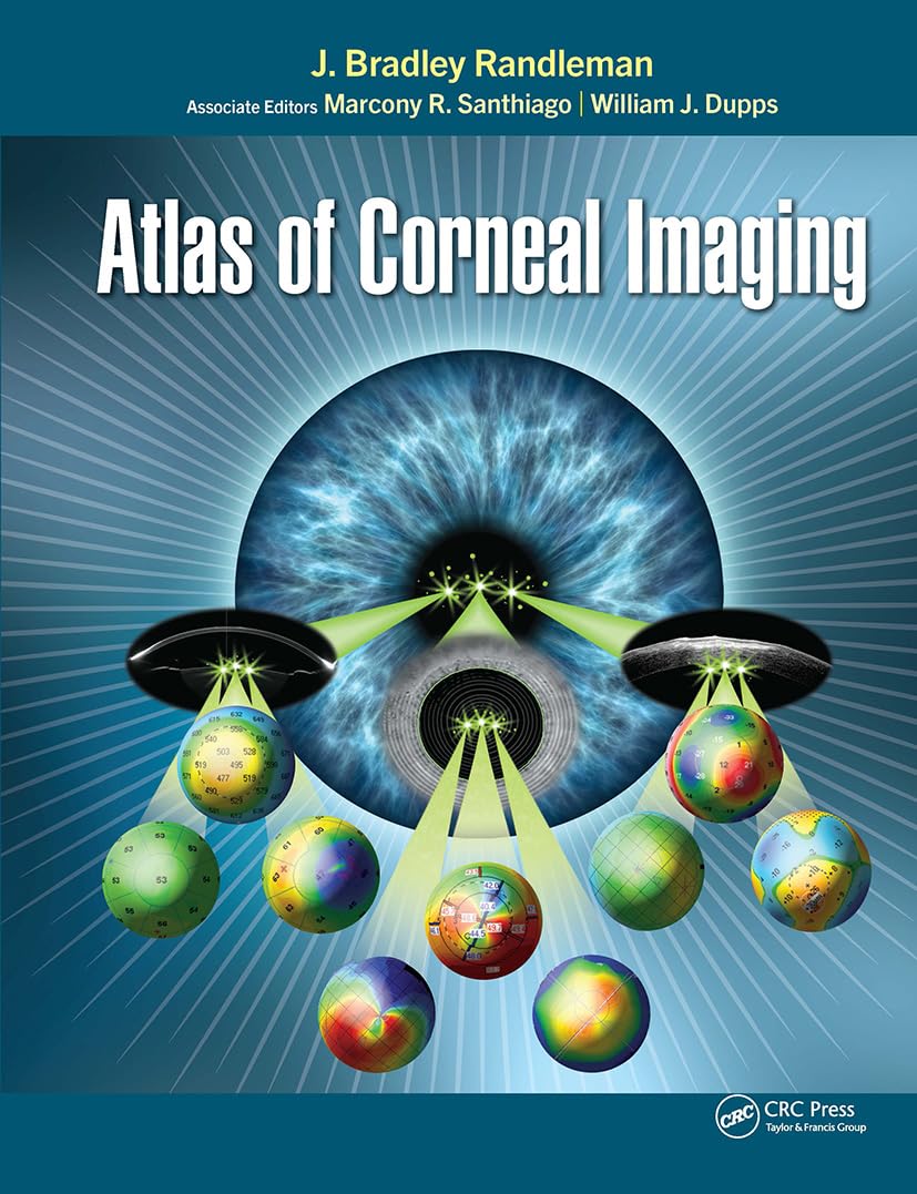
Atlas of Corneal Imaging is a comprehensive reference tailored for physicians, surgeons, and trainees seeking in-depth insights into corneal imaging. This indispensable resource, expertly crafted by Drs. J. Bradley Randleman, Marcony Santhiago, and William J. Dupps Jr, encompasses a wide array of essential topics and cutting-edge techniques in the field. With a wealth of over 1200 illustrative images and figures, this atlas offers a detailed exploration from basic map interpretation to advanced diagnostic applications.
By guiding readers through the intricate process of analyzing corneal images using diverse techniques and state-of-the-art technologies, this atlas offers a holistic view of the cornea’s fundamental methods and pathological processes. Practitioners can directly visualize how different devices would display various pathologies, aiding in the recognition of subtle findings and signs of weakening or pathology across different presentations and imaging devices.
Key features of the Atlas of Corneal Imaging include:
– Topographic patterns and mapping
– Evaluations of corneal ectasia
– Assessments of cornea and refractive surgery
– Clinical correlations with corneal disorders
– Complications related to cornea and refractive surgery
– Evaluation for cataract surgery
This resource serves as a vital tool for practitioners, bridging the gap in available corneal imaging references by adopting an image-first approach to understanding the myriad technologies used for imaging the cornea. With a focus on clarity and comprehensive coverage, this atlas empowers readers to enhance their diagnostic capabilities and deepen their understanding of corneal imaging techniques.
Explore the Atlas of Corneal Imaging to unlock a treasure trove of knowledge and visual insights that will enrich your practice and elevate your expertise in cor
Authors:
J. Bradley Randleman (Author)
Edition:
1st
Publication Date:
July 15, 2022
From the book:










Reviews
There are no reviews yet.