
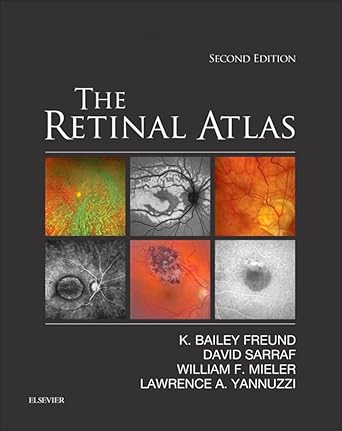
The Retinal Atlas, 2nd Edition, is your essential reference guide to retinal diseases, offering over 5,000 images and comprehensive illustrations of the entire spectrum of vitreous, retina, and macula disorders. As a trusted resource for retina specialists, comprehensive ophthalmologists, and ophthalmology residents and fellows, this updated edition provides definitive insights into this rapidly advancing field of retinal research and imaging.
Authored by renowned experts Drs. K. Bailey Freund, David Sarraf, William F. Mieler, and Lawrence A. Yannuzzi, the 2nd Edition of The Retinal Atlas includes:
– A complete visual guide to advanced retinal imaging and diagnosis, covering early and later stages of retinal diseases.
– Enhanced understanding of disease patterns with comparison imaging modalities, high-power views, panoramic disease visuals, and magnified views of key areas.
– User-friendly design featuring color coding for different imaging techniques, arrows, labels, and magnified images highlighting key lesions and intricacies.
– In-depth coverage of all current retinal imaging methods including: Optical Coherence Tomography (OCT), Indocyanine Green Angiography, Fluorescein Angiography, and Fundus Autofluorescence.
– Insight into expanding OCT uses such as Spectral Domain and En Face OCT, and evolving retinal imaging modalities like ultra-wide-field fundus photography, angiography, and autofluorescence.
– Extensive library of common and rare case illustrations contributed by a select team of international leaders in retinal imaging.
In addition, this award-winning title offers new images with multimodal illustrations, expanded coverage on key topics, and new disorders and classifications, making it the most comprehensive atlas of
Authors:
K. Bailey Freund (Author),
(Author),
(Author),
(Author)
Edition:
2nd
Publication Date:
November 14, 2016
From the book:


Reviews
There are no reviews yet.